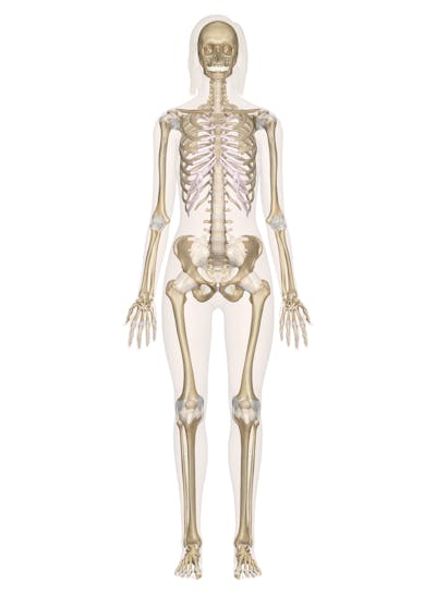
Back Human Bones Labeled : Body Cavity Diagram Koibana Info Body Bones Human Body Anatomy Human Bones Anatomy : The human skeleton anatomy chart shows three views of the human skeleton (front, back and side) and is painstakingly labeled and painted, producing one of the most captivating and beautiful anatomical charts available.
Back Human Bones Labeled : Body Cavity Diagram Koibana Info Body Bones Human Body Anatomy Human Bones Anatomy : The human skeleton anatomy chart shows three views of the human skeleton (front, back and side) and is painstakingly labeled and painted, producing one of the most captivating and beautiful anatomical charts available.. Bones of the pelvis and lower back the bones of the pelvis and lower back work together to support the body's weight, anchor the abdominal and hip muscles, and protect the delicate vital organs of the vertebral and abdominopelvic cavities. The red lines point individual bones and the names are writen in singular, the blue lines conect to group of bones and are in plural form. The axial skeleton is made up of the skull, backbone, breastbone, and ribs. Spinal anatomy and back pain. Overview of bones & the axial skeleton.
By adulthood, it is a large, triangular bone, that forms the base of the. The patella or the kneecap is the thick triangular bone of the knee. See human back anatomy stock video clips. Arms and hands bones names. Bones are often thought of as static structures which only offer structural support.
4 196 Skeletal System Vector Images Free Royalty Free Skeletal System Vectors Depositphotos from st2.depositphotos.com The skull also provides housing for the eye and optic nerve. The most common variations include sutural (wormian) bones, which are located along the sutural lines on the back of the skull, and sesamoid bones which develop within some tendons, mainly in the hands and feet. Using this atlas of human anatomy of the spine and back. Some individuals may also have additional (i.e., supernumerary) cervical ribs or lumbar vertebrae. The vertebral column of the lower back includes the five lumbar vertebrae, the sacrum, and the coccyx. The femur or the thigh bone is closest to the body. Make you have what you looking for. Bones are classified by their shape—as long, short, flat, and irregular.
It is a part of the hip and the knee.
Spinal anatomy and back pain. Human body muscles human body organs human body parts human organ diagram body organs diagram anatomy organs anatomy bones heart anatomy body muscle anatomy. Vertebrae, bones, joints, ligaments, muscles, muscular system, fascia, arteries, veins, nerves and various adjacent organs. They also provide for the attachment of muscles, and help us move around. To learn all about the skeleton system in the human body, check out this guide. This anatomy chart is ideal for higher education or patient consultation. Human back bone chart, find out more about human back bone chart. The human skeleton anatomy chart shows three views of the human skeleton (front, back and side) and is painstakingly labeled and painted, producing one of the most captivating and beautiful anatomical charts available. All the other bones in the skull are firmly attached to one another by. Your skeleton can be divided into two main parts. See human back anatomy stock video clips. Flexibility especially in the lower back and neck allowing us to bend and twist in a full variety of movements strength provided by the bones discs joints and supportive muscles and connective tissue that allows us to stand upright and move about with precision. The axial skeleton is made up of the skull, backbone, breastbone, and ribs.
Your skeleton can be divided into two main parts. The bottom of the spine is called the sacrum. The most common variations include sutural (wormian) bones, which are located along the sutural lines on the back of the skull, and sesamoid bones which develop within some tendons, mainly in the hands and feet. No need to register, buy now! The bones of the superior portion of the skull are known as the cranium and protect the brain from damage.
Bbc Science Nature Human Body And Mind Anatomy Skeletal Anatomy from www.bbc.co.uk This includes the head, facial, hyoid, auditory, trunk, ribs, and sternum. The longest and the strongest bone in the human skeletal system as you can observe in the labeled skeleton diagram of the human body. Flexibility especially in the lower back and neck allowing us to bend and twist in a full variety of movements strength provided by the bones discs joints and supportive muscles and connective tissue that allows us to stand upright and move about with precision. Your skeleton can be divided into two main parts. Bones of the pelvis and lower back the bones of the pelvis and lower back work together to support the body's weight, anchor the abdominal and hip muscles, and protect the delicate vital organs of the vertebral and abdominopelvic cavities. However, they truly function as an organ. The bones of the skull. Arms and hands bones names.
When most people mention their back, what they are actually referring to is their spine.
Your skeleton can be divided into two main parts. Vertebrae, bones, joints, ligaments, muscles, muscular system, fascia, arteries, veins, nerves and various adjacent organs. Arms and hands bones names. The most common variations include sutural (wormian) bones, which are located along the sutural lines on the back of the skull, and sesamoid bones which develop within some tendons, mainly in the hands and feet. Find the perfect anatomy rear view back human stock photo. That is the joint connecting the lower jaw, or mandible, to the rest of the skull. Bones are often thought of as static structures which only offer structural support. The remaining small bones or ossicles below the sacrum are also fused together and called the tailbone or coccyx. Flexibility especially in the lower back and neck allowing us to bend and twist in a full variety of movements strength provided by the bones discs joints and supportive muscles and connective tissue that allows us to stand upright and move about with precision. There is only one movable joint in the skull. To learn all about the skeleton system in the human body, check out this guide. It runs down the centre of the body. Diagram of a human female skeleton, back view.
The labeled human skeleton system is comprised of 206 different bones of various sizes and shapes, all with the primary purpose of providing support, protection, and shape to the human body. It is a part of the hip and the knee. By adulthood, it is a large, triangular bone, that forms the base of the. The number of bones in the human body at birth is 300. It is designed to be incredibly strong, protecting the highly sensitive nerve roots, yet highly flexible, providing for mobility on many different planes.
Skeletal System Labeled Diagrams Of The Human Skeleton from innerbody.imgix.net Diagram of a human female skeleton, back view. Bones are classified by their shape—as long, short, flat, and irregular. The muscles of the lower back help stabilize, rotate, flex, and extend the spinal column, which is a bony tower of 24 vertebrae that gives the body structure and houses the spinal cord. By adulthood, it is a large, triangular bone, that forms the base of the. Spinal anatomy and back pain. However, as a child grows, some of the bones fuse together. They also provide for the attachment of muscles, and help us move around. This includes the head, facial, hyoid, auditory, trunk, ribs, and sternum.
See human back anatomy stock video clips.
This includes the head, facial, hyoid, auditory, trunk, ribs, and sternum. The axial skeleton is made up of the skull, backbone, breastbone, and ribs. Beside that, we also come with more related ideas such labeled human skeleton diagram back, skull bones worksheet and skeletal system matching worksheet. The part of the nerve that emerges out of the spine is called the nerve root. That is the joint connecting the lower jaw, or mandible, to the rest of the skull. The vertebral column of the lower back includes the five lumbar vertebrae, the sacrum, and the coccyx. The spine anatomy is a complex structure. Primarily, they are referred to as long or short. On anatomical parts the user can choose to display the various structures in colored illustrations of the anatomy of the back and spine: The bones of the superior portion of the skull are known as the cranium and protect the brain from damage. Human back bone chart, find out more about human back bone chart. It is made up of several vertebral bodies usually fused together as one. No need to register, buy now!


0 Komentar